There are several tests to ensure that a woman is ovulating, and it is good practice for doctors to use more than one. The levels of hormones in the blood can be measured during at various stages of a menstrual cycle, which can help to pinpoint an abnormality that may affect ovulation; other blood tests may help identify how soon a woman may run out eggs. Measurement of progesterone, oestrogen, LH (luteinizing hormone), FSH (follicle-stimulating hormone), testosterone (which women also produce normally), prolactin, and, more recently, AMH (antimullerian hormone) levels are the most valuable.
Some women are worried that they have a high prolactin level, but if their periods are regular and if progesterone levels are normal and consistent with ovulation, then this is irrelevant. More commonly high prolactin levels are an indication that there may be polycystic ovaries. Occasionally, a very high level of prolactin can indicate a rare tumour of the pituitary gland.
The cells in a follicle that has just ruptured and released an egg stop producing the hormone oestrogen and start to release progesterone. This hormone, produced in relatively large amounts, prepares the lining of the uterus to receive any embryo that enters the uterus, help it to implant and to provide a suitable environment for pregnancy. A blood test can provide indirect evidence that ovulation has taken place. Expressed in the UK as ‘nmol (nanomoles) per litre’ and the peak level if ovulation occurs is about 30nmol per litre usually about a week before the next menstrual period is due.
Although the most widely used, progesterone tests are not foolproof. First, it is easy to mistime when the blood is taken. The level of progesterone is at its highest about a week after ovulation and stays high for three to five days. It is usual to take a blood test on the 21st day of the menstrual cycle, roughly seven days before the next period starts. But if a woman’s cycles are irregular or a period is a bit early or late, it is easy to miss the peak level. Some clinics repeat the blood test two or three days after day 21 if a longer cycle is anticipated. The level falls sharply immediately before a period, so a test taken within a day or two of bleeding may be meaningless. Many couples become discouraged when they have a low reading, but it may simply mean that the first day of the period occurred sooner or later than expected.
Most women have a slight rise in body temperature shortly after ovulation (probably because the higher level of progesterone increases metabolism). Charting body temperature is no longer regarded as essential, but I have included it because a few clinics suggest it, as it can provide a limited indication on whether a woman is ovulating. Recording temperature daily also allows the woman to take some charge in understanding her own cycle. However, many normally ovulating women have no discernible change in temperature. So perfectly fertile women who study their temperature chart may be caused needless anxiety, and others who are not ovulating may notice a rise in temperature after the mid-cycle and wrongly conclude that their ovaries are working properly. Some women believe that their temperature chart will tell them when they are at their most fertile and use it to time intercourse. The evidence for the value of timing like this is limited.
This hormone that has been known about since the 1940s and blood tests that assess AMH levels are used as a predictor of female fertility*.
Follicles that remain gradually develop to what is called the pre-antral stage, and the cells that line each follicle produce AMH. Measuring blood levels of AMH can give an indication of how many slowly maturing follicles remain in each ovary, and therefore how soon a woman might run out of eggs – the theory is, the more follicles the higher the level of AMH. Women age at different rates, so the loss of maturing follicles, and therefore eggs, varies too.
There are disadvantages. A woman’s AMH levels fluctuate and they can vary from one woman to another. In addition, how the levels are measured varies from one laboratory to another; the method used for detecting this hormone is not ideal, and not as yet well standardized – even the way the blood is stored can make a difference to the result. Nevertheless, AMH levels give an indication of how many eggs are likely to be collected during a single IVF treatment cycle. However, some women with quite high results do not produce many eggs, while others with low levels are relatively fertile. Measuring AMH level is controversial because it does not necessarily predict the chances of a pregnancy following IVF as women with quite low levels do occasionally get pregnant. High FSH levels can indicate a lack of ovarian reserve, but often the levels of FSH and AMH do not always correlate; a woman can have high FSH levels but normal AMH levels.
The advantage of the AMH blood test is that it can be done at any time during the cycle. It is used to predict, with modest success, the ovarian reserve after a cancer treatment, after surgery on the ovaries, and the chances of hyperstimulation if gonadotropins are used to stimulate the ovaries. AMH levels are often raised in people with PCOS, but equally, it may be less good at predicting ovarian reserve in those with PCOS.
Understanding AMH levels is confusing. For one thing, they are usually given in nanograms per litre but sometimes in picomoles, which give a higher reading. A single measurement is usually not enough to decide whether eggs are running out. In addition, results vary so much ask your specialist what the normal levels are in the clinic you are attending.
Follicles also produce two closely related hormones: Inhibin A and B. Inhibin A is produced by the follicle that is about to ovulate (dominant follicle) and the corpus luteum (the empty follicle after ovulation). Inhibin B is produced by developing follicles, well before they are ready for ovulation. Inhibin B level varies during the menstrual cycle and is normally tested on day three when it is increasing rapidly. Thereafter the level starts to decrease many days before ovulation. At menopause, with the loss of follicles, inhibin A and B levels are very low or undetectable.
Inhibin B is the hormone that might help indicate ovarian reserve, but its use is still experimental. As ovarian reserve diminishes, the hormones used to stimulate ovulation during IVF do not work so well. Inhibin does give some indication of the number of eggs that remain in the ovary but, at present, levels of inhibin B do not seem that good predictor of likely response to IVF.
There are a number of tests that a specialist may undertake, ranging from an ultrasound scan to more invasive examinations that require a general anaesthetic.
An endometrial biopsy involves removing a small piece of the uterine lining (the endometrium) and examining it under the microscope. The biopsy is undertaken during the second half of the menstrual cycle to see whether uterine lining has been exposed and has responded to the progesterone that is normally produced by the ovary after ovulation. Endometrial biopsy has been used increasingly to look for biochemical markers that indicate whether the lining of the uterus is developing properly, and whether or not the uterus is capable of allowing an embryo to implant.
Endometrial biopsy is normally carried out between days 18 and 28 of a 28-day menstrual cycle. The cervix is examined and cleaned. A small pipe is inserted through the cervix and a tiny scraping of the uterine lining is removed. This can cause brief discomfort – a cramp-like period pain. Because some women are nervous about the pain that can occasionally occur many clinics do this biopsy when a patient comes for laparoscopy under general anaesthesia.
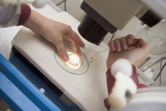
An ultrasound scan uses high-frequency sound waves that are passed through the body. The sounds are not audible, but when the sound waves hit tissue, echoes are given off, which are picked up by a receiver in the probe, analysed by a computer and displayed on a monitor. Sound waves are aimed at the ovaries through the abdominal wall or via the vagina, using a vaginal probe. The ovaries lie just behind the bladder and because water is a good conductor of sound, the best pictures may be obtained when the bladder is full. You may, therefore, be asked to drink water before the abdominal ultrasound, which can be uncomfortable. Very accurate pictures of the ovaries can also be produced with a small ultrasound probe in the vagina, avoids the discomfort of needing a full bladder.
An ultrasound scan can determine whether or not a follicle is growing. When ready to rupture and release an egg a mature follicle is approximately 2 centimetres. It can also detect a sudden decrease in size after ovulation, which can indicate that it has occurred.
Ultrasound is also an important technique for diagnosing polycystic ovaries, cysts and ovarian damage resulting from endometriosis and can be used to detect early pregnancy. This scan can also be used to assess fibroids in the uterus, but X-rays or MRI (magnetic resonance imaging) give a more detailed view. In recent years, ultrasound procedure called Hysterosalpingo-contrast-sonography, or HyCoSy has been used to assess whether the fallopian tubes are open and clear. A special saline solution and a contrast agent are introduced into the uterus, and can be observed flowing through the tubes. In my view X-ray and laparoscopy are much more accurate.
The Hysterosalpingogram, or HSG, is an X-ray of the uterus and fallopian tubes. It is an unduly neglected test that some doctors believe has been superseded by telescopic inspection (laparoscopy) under general anaesthetic. HSG provides information that it is difficult to obtain any other way, it is easy to do and less expensive than procedures that require an anaesthetic.
The X-ray is done following an internal examination and the procedure takes about ten minutes. A thin tube no larger than a ballpoint pen refill is passed through the cervix and a small amount of dye is injected into the uterus. The dye’s progress is monitored on a television screen and about six X-rays images may be taken. The quality of the shadow on the X-ray can give a clear outline of the inside of the uterus, and shadows inside the tubes can reveal whether the fallopian tubes are blocked. If digital X-ray equipment is used, adhesions inside the uterus, fibroids, scarring of the uterine muscle and polyps, even congenital abnormalities of the uterus can be seen on and HSG image. In the area where the tubes join the uterus, the ‘plumbing’ is extremely delicate and small, a good HSG image will reveal scar tissue or polyps in the tube itself, even scarring in the tubal lining and its folds, more effectively than a laparoscopy. This is invaluable because often there is tubal disease even though the tubes are not blocked; this kind of scarring increases the risk an ectopic pregnancy.
Many clinics request an HSG as soon as possible after the first visit to the clinic. By the time, a couple attends their second appointment there will be a detailed assessment of the quality of the tubes and the uterus. HSG used to have a reputation for causing discomfort, but most women do not even realise that the test has started or finished. Sometimes the insertion of the tube can be uncomfortable, but it is no worse than a period pain. Persistent pain after the HSG can indicate infection and is not normal. Any woman who experiences this should contact the hospital immediately.
I have seen many women after unsuccessful IVF in other clinics who have been told they have ‘unexplained infertility’. All too often they were told HSG was not necessary, or it was done inadequately. When we perform HSG on these patients we often uncover a clear reason for the failure of the earlier IVF attempts. As most of these contributing factors are correctable, it is a great pity that more emphasis is not placed on getting good quality X-rays before IVF treatment is started.
This is a medical imaging technique that uses strong magnetic fields and radio waves to produce detailed images of the body. The magnetic waves are produced by a large coil in a cylinder that surrounds the body. Moreover, because it does not use damaging X-radiation, it is entirely risk-free. It can be a slightly unnerving experience because the machine itself makes a considerable noise while it is operation and you have to lie very still for several minutes at a time inside the machine while images are displayed on a remote screen in another room. MRI gives very high-quality images, far better than those achieved, for example, using ultrasound, but not as a good as a high-quality X-ray.
MRI is an expensive procedure, and probably not used often enough in cases of infertility. It can, however, provide extremely valuable information. We use it when we suspect that the uterine wall may be abnormal. In particular, it can provide evidence of damage to the wall of the uterus and scar tissue in the muscle. We use it when we suspect that the patient has the condition called adenomyosis* and occasionally to investigate cysts in the pelvis. These are frequent causes of infertility and may also cause painful periods. It is quite common in older women who have been unsuccessful with IVF.
This is by far the most important single test for female infertility. In my view it, laparoscopy should nearly always be considered before entering an IVF programme unless it is known that a woman has no fallopian tubes, or that there is no possibility of corrective surgery.
Laparoscopy needs to be done under general anaesthetic in an operating theatre, but can normally be done as a day-case. Some clinics perform it under a local anaesthetic, but a general anaesthesia allows the surgeon to make a more detailed inspection.
A thin telescope is inserted into the abdominal cavity through a small hole made in the navel. Carbon dioxide, passed into the abdomen, separates the organs so that they can be viewed more easily. The telescope is no thicker than a fountain pen, but with the improvement in modern optics, superb-quality photographs can be taken. A surgeon can inspect the outside of the uterus, he or she can inject dye through the tubes to see if they are open. An endometrial biopsy may be done at the same time. The whole procedure may take anything from 15 to 30 minutes, or longer if it is being used for keyhole surgery (for example to release the fallopian tubes from adhesions). After laparoscopy more women immediately conceive than would be expected by chance. Historically, up to about 15 per cent of our patients with open tubes conceive within three months of laparoscopy
What to expect after laparoscopy
There will normally be two small dressings on the abdomen: one covers a single stitch in the navel, and the other a tiny hole near the pubic hairline. This second hole was used to place any fine probes shaped like small knitting needles into the abdominal cavity to move tissues around to get a better view. Laparoscopy usually causes very little pain or discomfort although some women may feel unwell and need to rest in bed for 24 hours. The most common side effects are:
Benefits of laparoscopy:
A small telescope, called a hysteroscope, is passed into the uterus through the vagina, normally under a short general anaesthetic on a day visit to the hospital. It is an excellent means of detecting any polyps, uterine fibroids, adhesions or congenital abnormalities, which may be suspected following the results of an HSG X-ray. It can also be used to treat some of the found after HSG conditions by guiding instruments inside the uterus.
Tuboscopy requires a fine telescope to be inserted through the abdominal wall under general anaesthetic, in order to inspect the inside of the ovarian end of the fallopian tube. This test, which can be combined with laparoscopy, above, is of only limited value and can be very expensive. An alternative is a falloscopy, which involves passing a very fine telescope connected to a television camera in the operating theatre, into the fallopian tube, via the vagina, cervix and uterus. This telescope is about the thickness of a piece of linen thread and gives the surgeon a view of the uterine end of the fallopian tube. However, the optic fibre is so narrow the resolution of the picture is not as good as that seen using tuboscopy. It probably has even less value that tuboscopy and is only included here for completeness. Both these tests straightforward, but less useful to the patient except perhaps when assessing the pelvis of a woman who has suffered an ectopic pregnancy.
Despite countless breakthroughs in medical science, we still do not understand why some pregnancies will end in tragedy. For most of us, having a child of our own is the most fulfilling experience of our lives. All of us can imagine the desperation and sadness of parents who lose a baby, and the life-shattering impact that a disabled or seriously ill child has on a family.
Professor Robert Winston’s Genesis Research Trust raises money for the largest UK-based collection of scientists and clinicians who are researching the causes and cures for conditions that affect the health of women and babies.
Essential Parent is proud to support their wonderful work. You can learn more about them here.